Apollo, a healthy neutered male 9-month-old Siberian husky, was walking on a leash in Ken Caryl, when he and his owner spotted a rattlesnake on the sidewalk. Apollo was allowed to investigate. The rattlesnake struck Apollo in the eye. He was immediately taken to an emergency clinic where a severe corneal injury with severe ocular pain was diagnosed and treated with topical antibiotics, antivenin, carprofen, and tramadol. Given the severity and nature of the ocular injury, enucleation was recommended to relieve the ocular pain from what would likely be a blind eye. The client refused eye removal and requested referral to a specialist. Here is what I saw:
Bean
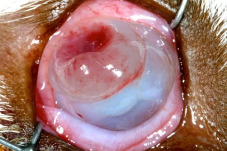
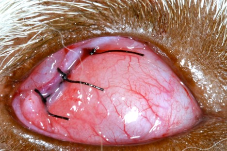
2 days following a witnessed rattlesnake bite to the right eye. Severe extensive keratomalacia attributed to the proteinases in the venom. Poorly visualized intraocular structures. Severe blepharospasm. Given the likelihood of catastrophic corneal perforation and the absence of healthy cornea to receive a graft, I placed a third eyelid flap sutured to the superior bulbar conjunctiva with three 5-0 silk sutures in a horizontal mattress pattern. An Elizabethan collar and topical ofloxacin QID and carprofen 2.2 mg/kg PO BID were prescribed.
Bean
Ten days later, I removed the sutures that retained the third eyelid flap, and here is what I saw:
Bean
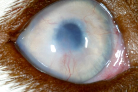
Complete keratomalacia with anterior bulging in some areas and corneal rarefaction in other areas. Early 360-degree deep corneal neovascularization. Absent menace response. Intact dazzle reflex. Eye open and apparent comfortable.

3 weeks post-injury:

Bean
Progressive 360-degree deep corneal neovascularization. Still unable to visualize the intraocular structures. Intact menace response and dazzle reflex.

Six weeks post-injury:

Bean
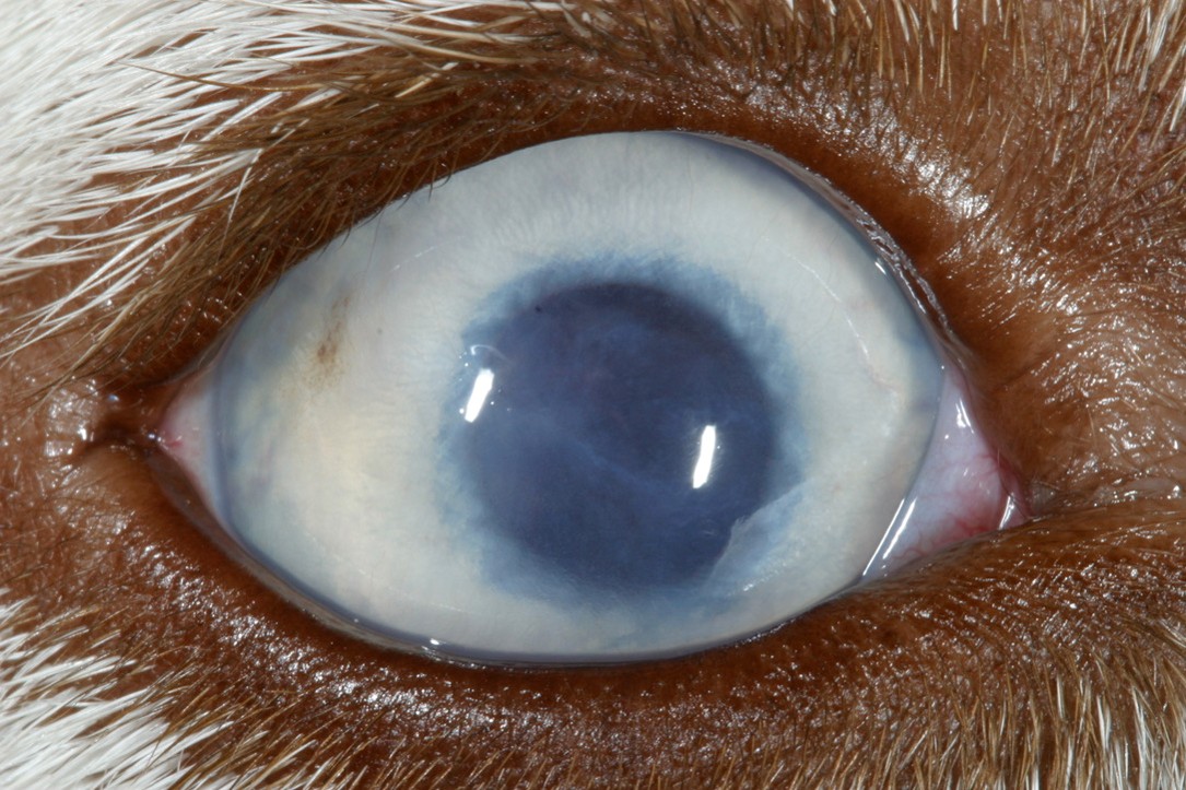
Resolving corneal neovascularization. Remodeled cornea to normal thickness with moderate corneal stromal fibrosis. No uveitis. A healthy retina and optic disk was visbible. The eye was visual with intact tracking of thrown cotton balls.

3 months post-injury:

Bean
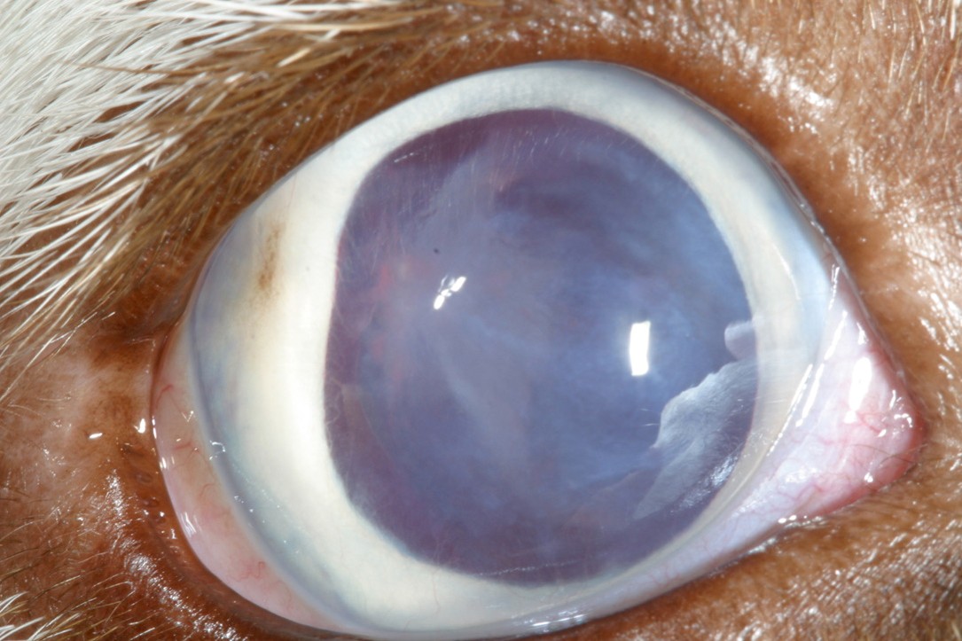
3 months post-injury after pupillary dilation with tropicamide:
Bean
The eye was visual and comfortable with mild diffuse corneal fibrosis, two focal temporal corneal scars and subjacent focal posterior synechia and focal temporal cataract.

At the last examination 4 years post-injury, the remained visual and comfortable with nonprogressive cataract.

Key Takeaways:

1. Enucleation will always solve a severe eye problem. However, sometimes supporting an eye and allowing it to heal will result in a comfortable eye and sometimes vision. Sometimes, patience is warranted. This is especially true in young patients like Apollo.

2.We commonly repair serious corneal defects by grafting with collagen grafts, conjunctiva, and autologous cornea. These procedures require advanced microsurgical training and specialized equipment including an operating microscope. A third eyelid flap is an unsophisticated procedure that can be placed with minimal training and materials and equipment that are readily available in veterinary practices. Sometimes, amazing healing can occur beneath a third eyelid flap.