
Pigmentary Keratitis is a common, progressive, and potentially blinding ocular disease in pugs, with reported prevalence rates as high as 96%. The condition is characterized by corneal pigmentation that typically originates at the nasal limbus and gradually advances across the cornea. Vision becomes impaired when pigmentation extends into the visual axis, directly overlying the pupil. Disease severity varies widely, but pugs younger than five years of age with involvement of the visual axis are at the greatest risk of progressive vision loss and eventual blindness. Management generally requires long-term topical immunomodulatory therapy with tacrolimus or cyclosporine. Although topical corticosteroids can be effective, their use is limited by the increased risk of opportunistic bacterial keratitis in a breed predisposed to exposure-related corneal disease due to lagophthalmos and exophthalmos.
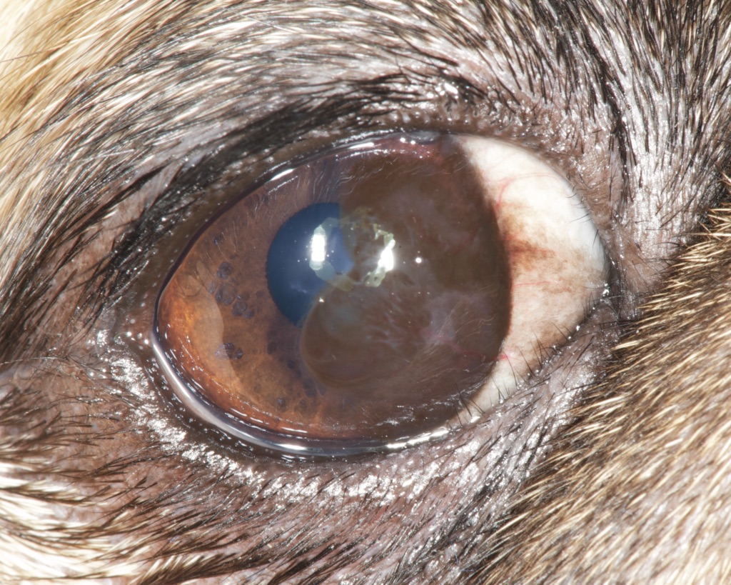
Moderately advanced corneal pigmentation with partial obstruction of the visual axis]
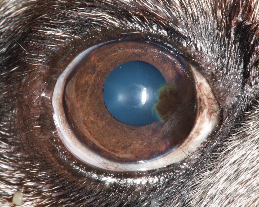
Early corneal pigmentation with no impact on vision
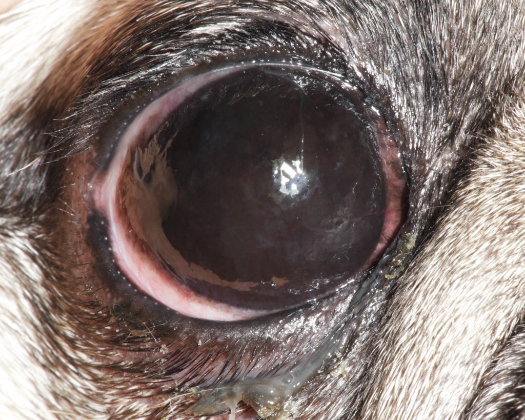
Complete corneal pigmentation and severe vision impairment
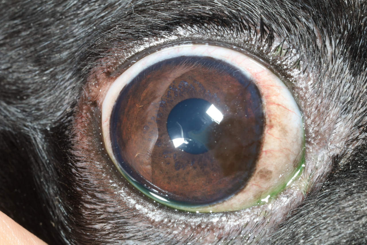
Vision threatening corneal pigmentation in a 3 year old Pug
Recent studies have demonstrated that medial canthoplasty is a safe and effective procedure for slowing or even reversing corneal pigmentation while helping preserve vision.1,2 The surgery involves removing the medial canthus, correcting localized entropion by everting the nasal lower eyelid, and reconstructing the nasal canthus to reduce dryness and mechanical irritation at the nasal limbus. Our experience mirrors the published findings, with treated pugs showing greater reductions in corneal pigmentation than those managed with medical therapy alone. This procedure is particularly appropriate for pugs whose pigmentation continues to progress despite consistent medical treatment. Please give us a call if you’d like to schedule or learn more about nasal canthoplasty.
1. Allgoewer, I. Long-term effects of various therapies for pigmentary keratitis in Pugs.
2. Allgoewer, I. Simplified medial canthoplasty: Technique and early postoperative complications in 601 dogs (1180 eyes).
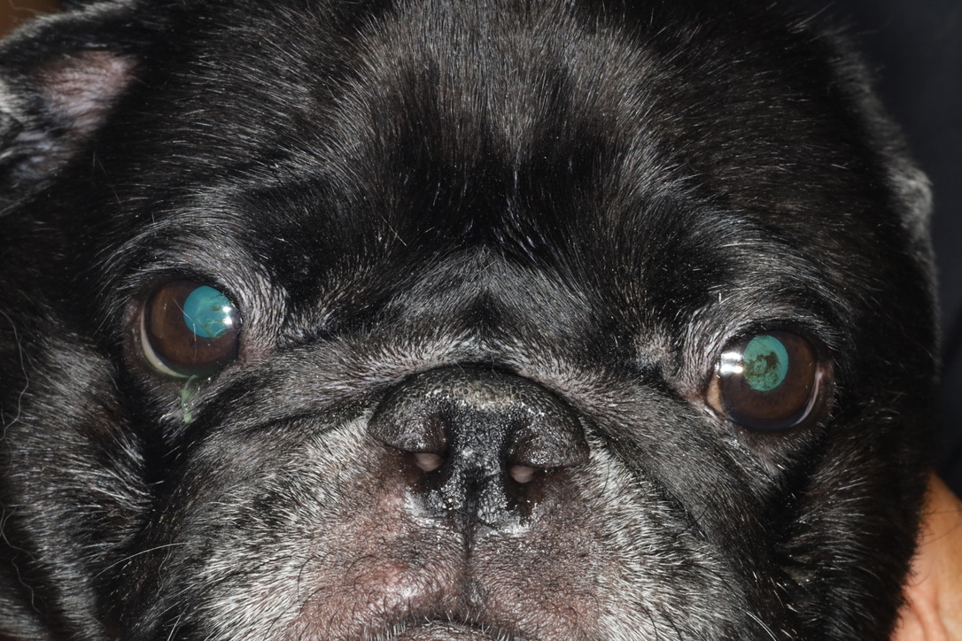
Patient 6 months after nasal canthoplasty.
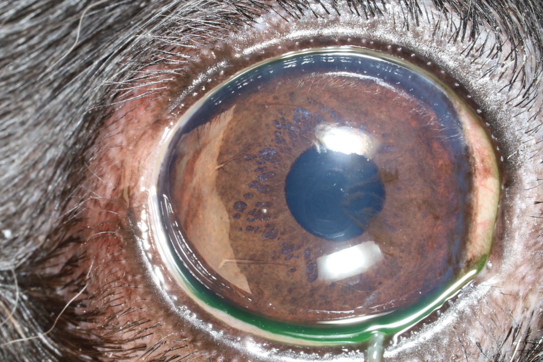
Rarefied corneal pigmentation and reduced sclera visible 6 months after nasal canthoplasty.